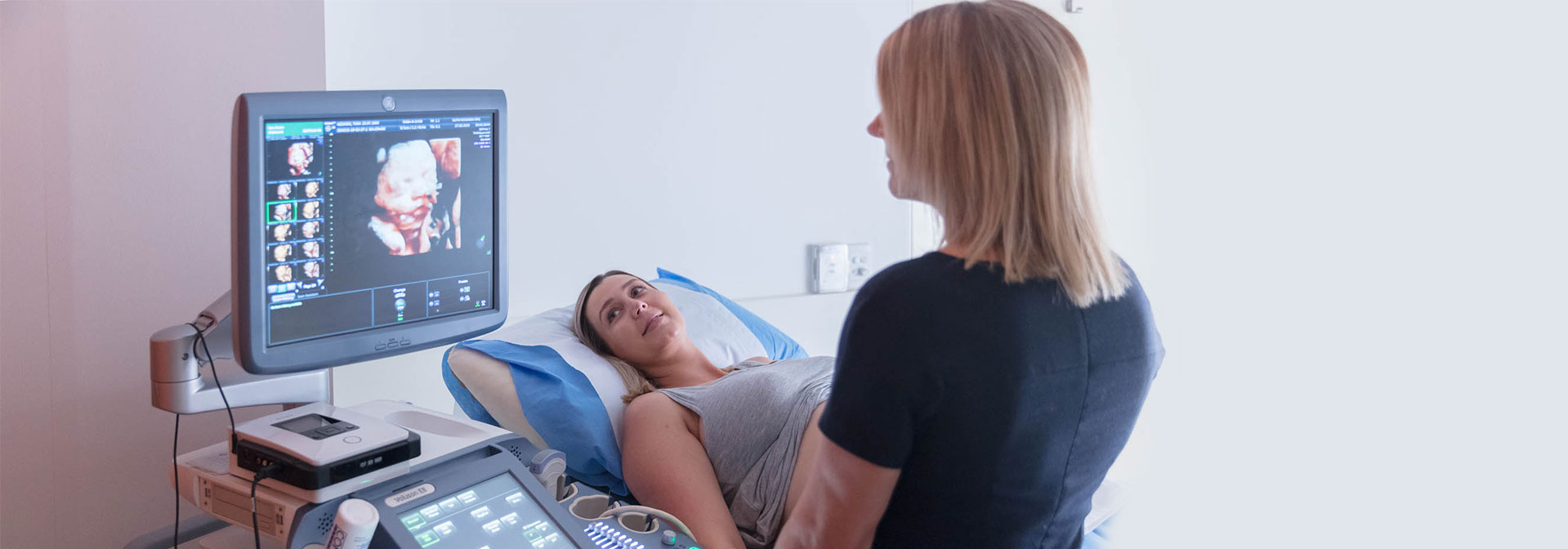
We recommend your First Trimester Ultrasound is performed at 13 weeks, we call this your anatomy scan, at this time you will be provided with information that has not yet been available during your pregnancy, which is why the 13 week scan is important.
We will check that your baby is growing well and confirm your due date along with some other key checks including:
- Anatomy of your baby
- Nuchal Translucency
- Pre-eclampsia risk
- Placenta
- Your ovaries
At 13 weeks, the anatomy of your baby can be assessed in great detail. Technology has advanced significantly and we can now recognise or suspect any structural abnormalities at 13 weeks, these checks are best identified via an internal ultrasound (ideally performed between 12 weeks 5 days and 13 weeks 2 days).
The nuchal translucency (NT) of the fetus is identified and measured during this narrow window of time. The NT is a collection of fluid between the skin and soft tissues of the neck, it is often increased with Down syndrome and other chromosomal or congenital abnormalities, you can read more about Screening for Down syndrome here.
We will measure your uterine artery blood flow, combined with other maternal factors for a pre-eclampsia risk assessment. Pre-eclampsia is a disease affecting the health of both mother and baby, it is one of the primary reasons that you may need to deliver your baby earlier, you can read more about pre-eclampsia here.
During this scan, we will look at the insertion of the cord into the placenta. The proportion of amniotic fluid to baby size is greater at this early stage, which allows for good visualization of the cord and therefore helps to detect if there is any variation from normal, which we can manage throughout your pregnancy.
We will also assess your ovaries, as your pregnancy progresses and the womb fills the pelvis, your ovaries are repositioned and often impossible to locate with an ultrasound. The 13 week ultrasound is therefore an opportune time to perform an assessment of your ovaries.
How is the scan performed?
Our protocol is to perform an internal ultrasound at the 13 week anatomy scan. In the majority of cases, this provides superior image quality and therefore finely detailed assessment of your baby.
What is the risk of the scan?
Both internal and transabdominal ultrasound examinations are safe throughout pregnancy.


